X-ray examination
The dentist cannot check all spots of the oral cavity with a mirror and probe. To refine an oral examination, to explain certain symptoms or to follow up a specific treatment, the dentist takes an X-ray.

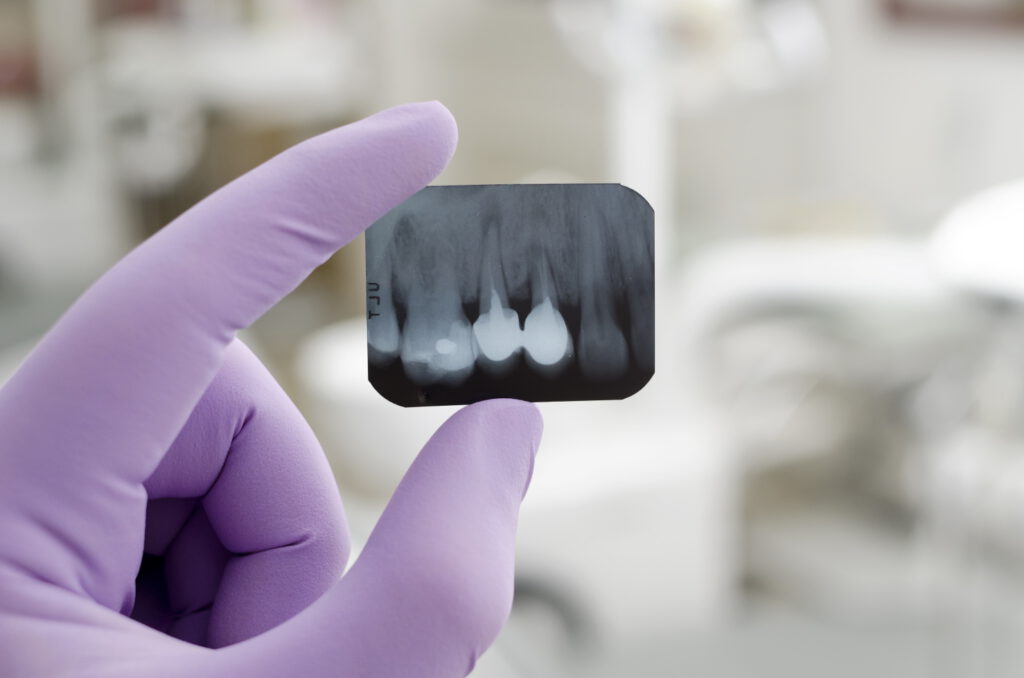
X-rays are pictures taken using X-rays. X-ray waves penetrate to a greater or lesser extent through parts of the human body, such as bones and muscles. Therefore, X-rays can be used to get a picture of the internal structure of the human body. There are different sizes of photos and depending on what the dentist wants to check, he/she will decide what kind of photo to take.
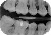
An intra-oral photo: a small photo or plate is placed in the mouth at the height of the tooth(s) and the environment that the dentist wants to view (bite-wing photo, periapical (root) image or solo photo)


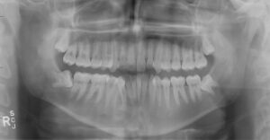
A panoramic view (OPG): a large plate rotates around the patient's face; in this photo the entire upper and lower jaw, the jaw joint and the bone can be viewed
An X-ray provides additional information about the teeth and jawbone that is not obtained with the naked eye. This allows the dentist to make a choice between the need for treatment or, if possible, following the disease process (monitoring). This reduces the risk of unwanted damage due to a progressive disease process (eg tooth decay or loss of bone tissue) or unnecessary treatment.
Why does the dentist take X-rays?
The dentist examines the mouth with a mirror and a probe (this is the 'bracket'). However, this allows the dentist to only see the outside of the teeth and gums. Sometimes it is also necessary to look 'into' the molars and the jawbone. For example, because the dentist suspects that there are cavities under a filling or crown. But also to check whether an implant fits properly or whether a root canal treatment is going well or has been successful.
The dentist can also see whether all teeth – which have yet to erupt in children – are present. X-rays are also used to view the position of the roots of teeth, the health of the jawbone and whether any remains of tooth roots have been left behind in the jaw.
Risks X-rays
Everyone is exposed to background radiation from, for example, the earth and the universe on a daily basis. The amount of radiation to which we are exposed differs per area. In the Netherlands we receive less radiation than in, for example, high-lying and mountainous places. A minimal amount of radiation is used when taking X-rays at the dentist. This minimizes the risk of adverse health effects. The amount of radiation, for example, can be compared with the amount of radiation you incur during a 14-day winter sports holiday. Of course, with radiation, no matter how small, the usefulness of the X-ray must always be weighed up against the risks of radiation. There is then a chance that an inflammation or other dental abnormality will not be detected or will be detected too late.
X-rays during pregnancy
In dentistry, only X-rays are taken of the teeth or jaw. The radiation will therefore not come close to the unborn child. For this reason, dental X-rays during pregnancy can in principle be taken without objection. However, if your dentist is familiar with your pregnancy, he will only take pictures in the most necessary cases.
Alternative treatment
If it appears during oral examination or screening for tooth decay that additional information is desired or necessary, the dentist will take an X-ray. There is no alternative to this treatment.
Source: KNMT and Dr. GA van der Weijden, dentist periodontist/implantologist
Come visit us!
- info@mondzorg-valerius.nl
- +31 0 70 870 1466
- Valeriusstraat 25, 2517 HM The Hague